Pleural Mesothelioma Ct - Diagnosis Of Malignant Pleural Mesothelioma Stanford Health Care : However, here we report a rare case of mpm diagnosed in a healthy young male patient without significant asbestos exposure.
Pleural Mesothelioma Ct - Diagnosis Of Malignant Pleural Mesothelioma Stanford Health Care : However, here we report a rare case of mpm diagnosed in a healthy young male patient without significant asbestos exposure.. J comput assist tomogr 1983; Although, ct has intrinsic limitations due to low soft tissue contrast and the current staging system as well as criteria for evaluating response, it does not consider the complex growth pattern of this tumor. Staging is a way to describe the cancer and whether and how far it has spread beyond its original site. ct of malignant pleural mesothelioma. If that ct scan showed advanced disease, a further ct scan may not be necessary.
A case of malignant pleural mesothelioma with osseous and. ct manifestations in 50 cases. This retrospective study aimed to investigate the prognostic value of the suvmax in patients with mpm. pleural effusion occurred in 74% of those patients. Shiba n, kusumoto m, tsuta k, et al.
Giant Malignant Mesothelioma In The Upper Mediastinum A Case Report from www.spandidos-publications.com Metintas m, ucgun i, elbek o, et al. Findings that could indicate mesothelioma is a thickening of the pleura, deposits of calcium on the pleura, fluid in the space between the chest wall and lungs, and changes in the lungs themselves from exposure to asbestos. Staging of malignant pleural mesothelioma: Asbestosis is often detected through ct scans. pleural mesothelioma is a type of cancer caused by inhaling asbestos fibers. ct scans typically take 10 to 15 minutes to perform. Comparison of ct and mr imaging. The 2016 mesothelioma audit data reported that in the uk in 2014 pleural mesothelioma accounted for 2179 cases (97%), with 70 peritoneal cases (approximately 3%).1 in 2007, the british thoracic society (bts) statement on mesothelioma was published in response to a request from the
The main test to stage mesothelioma is a ct scan.
J comput assist tomogr 1983; ct scans are the primary imaging technique for a patient with suspected pleural mesothelioma. These fibers get lodged into the protective lining of the lungs (the pleura), causing genetic mutations in the surrounding cells. The purpose was to assess the accuracy of ct scan based preoperatively measured tumor volume and thickness compared to actual tumor weight of resected mpm specimen and pathologically assessed tumor. Unlike other malignant mesotheliomas that occur in the abdomen, heart, or testicles, this form of mesothelioma is a cancer that occurs in the pleura, or the tissue lining of the chest cavity that encloses the lungs. A case of malignant pleural mesothelioma with osseous and. Your general practitioner (gp) will assess your symptoms. Malignant pleural mesothelioma (mpm) is a highly aggressive malignant tumor that arises from mesothelial cells of pleural cavity. Because mesothelioma may spread to the diaphragm, an mri may be used to look at the diaphragm, the muscle used for breathing, which separates the chest from the abdomen. 37 full pdfs related to this paper. The 2016 mesothelioma audit data reported that in the uk in 2014 pleural mesothelioma accounted for 2179 cases (97%), with 70 peritoneal cases (approximately 3%).1 in 2007, the british thoracic society (bts) statement on mesothelioma was published in response to a request from the European journal of nuclear medicine and molecular imaging, 2014. ct is the first imaging technique used for diagnosis, staging, and assessment of therapy response.
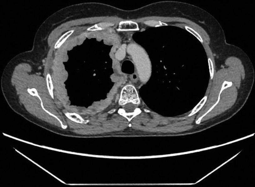
1 most cases of mpm are associated with a history of asbestos exposure, with an incubation period of 10 to 50 years or more. Although the chest film findings of pleural mesothelioma are well described, there are few descriptions of the findings of computed tomography (ct). Typical mediastinal pleural involvement with mesothelioma remains eligible; Evaluation with ct, mr imaging. Malignant pleural mesothelioma is a rare tumor.
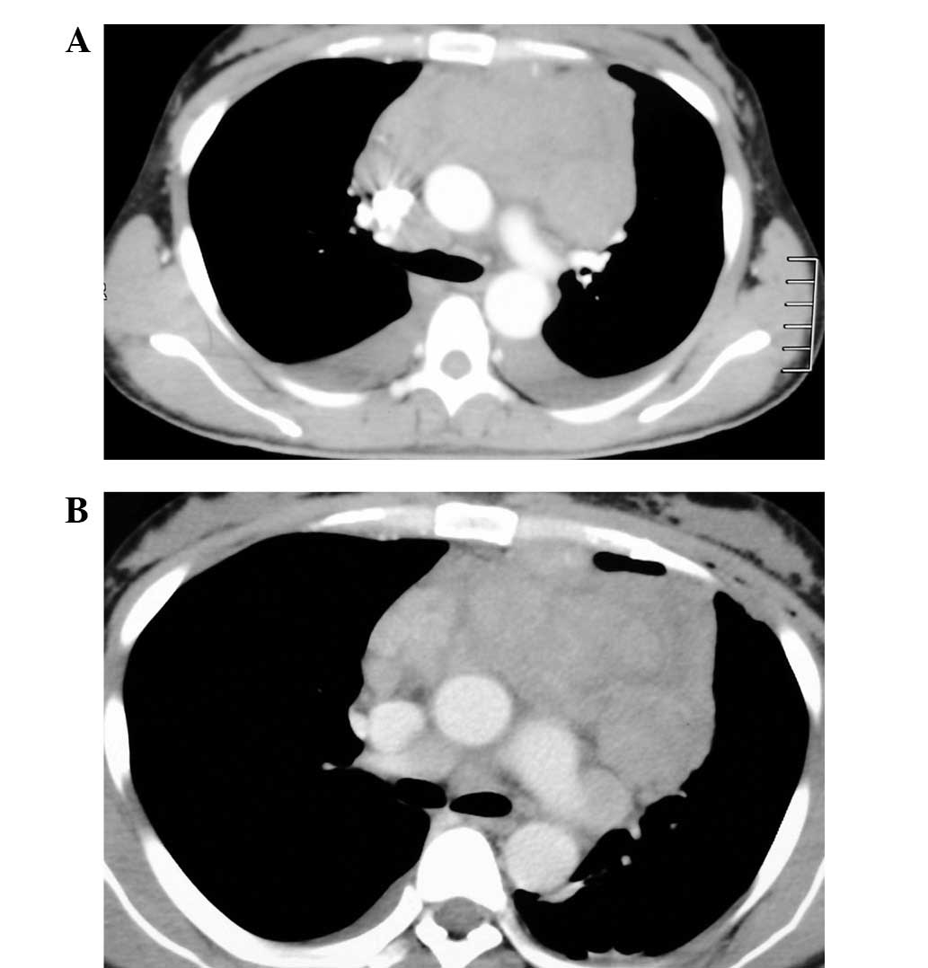
Tumor Regression In A Pleural Mesothelioma Patient Treated With Download Scientific Diagram from www.researchgate.net 37 full pdfs related to this paper. Malignant pleural mesothelioma (mpm) is a highly aggressive malignant tumor that arises from mesothelial cells of pleural cavity. However, here we report a rare case of mpm diagnosed in a healthy young male patient without significant asbestos exposure. pleural mesothelioma is the most common of the four mesothelioma types, making up approximately 75% of all cases diagnosed. Computerized tomography (ct scan) of the chest or abdominal region, along with magnetic resonance imaging or mri, and tissue biopsy of the tissue that makes up the lining of the lungs are some of the tests that a doctor usually runs to check for mesothelioma. Pleura) and chest wall (the "parietal" To asc plus mvp (four cycles of mitomycin 6 mg/m 2, vinblastine 6 mg/m 2 and cisplatin 50 mg/m 2 every 3 weeks (n=137)) or to asc plus vinorelbine (one injection of vinorelbine 30 mg/m 2. pleural mesothelioma affects the lining of the lungs (pleura) and is the most common form of this rare cancer.
mesothelioma represents a serious cancer associated with prior asbestos exposure.
Staging of malignant pleural mesothelioma: "the diagnostic performance of ct in routine clinical practice is insufficient to exclude or confirm pleural mesothelioma," ct scans typically take 10 to 15 minutes to perform. A ct (or cat) scan or an mri is usually performed. Your general practitioner (gp) will assess your symptoms. This stage describes patients whose tumors haven't started growing and spreading beyond their origin. Biopsies if imaging scans reveal signs of pleural mesothelioma, a pathologist will perform a biopsy, which is a procedure that removes a small fluid or tissue sample from a patient's body to test it for. These findings suggest pleural plaques are not common in the general public. These fibers get lodged into the protective lining of the lungs (the pleura), causing genetic mutations in the surrounding cells. In 13 of these 76 patients with pleural plaques, the original ct scan was performed due to suspicion of prior asbestos exposure. Your gp will conduct a physical examination and order tests. The main symptoms are shortness of breath, pain when breathing, chest/shoulder/upper arm pain, loss of appetite, weight loss, and persistent cough or bouts of pneumonia. The association between pleural plaques and pleural mesothelioma remains controversial.
"the diagnostic performance of ct in routine clinical practice is insufficient to exclude or confirm pleural mesothelioma," Centrally located tumors with radiographic evidence (ct or mri) of local invasion of major blood vessels; Shiba n, kusumoto m, tsuta k, et al. ct is the first imaging technique used for diagnosis, staging, and assessment of therapy response. Your gp will conduct a physical examination and order tests.
Measuring Malignant Pleural Mesothelioma Springerlink from media.springernature.com pleural mesothelioma affects the lining of the lungs (pleura) and is the most common form of this rare cancer. Malignant pleural mesothelioma (mpm) is an aggressive thoracic malignancy with a dismal prognosis. (9 days ago) increasingly, fdg pet/ct is used for diagnosis and management of malignant pleural mesothelioma. Pleura), as well as the diaphragm, and the heart and major blood vessels in the central portion of the chest. One study showed pleural thickening was evident in 94% of pleural mesothelioma patients who underwent a ct scan. Heelan rt, rusch vw, begg cb, et al. To asc plus mvp (four cycles of mitomycin 6 mg/m 2, vinblastine 6 mg/m 2 and cisplatin 50 mg/m 2 every 3 weeks (n=137)) or to asc plus vinorelbine (one injection of vinorelbine 30 mg/m 2. mesothelioma is often found after a person sees a doctor because of the symptoms they are having.
ct manifestations in 50 cases.
Computed tomography of malignant pleural mesothelioma. This report describes the ct findings in five cases of pleural mesothelioma. Staging is a way to describe the cancer and whether and how far it has spread beyond its original site. Malignant mesothelioma is a rare and insidious neoplasm with a poor prognosis. ct scans typically take 10 to 15 minutes to perform. In cases of peritoneal mesothelioma, fluid can build up in the abdomen (called ascites). "the diagnostic performance of ct in routine clinical practice is insufficient to exclude or confirm pleural mesothelioma," The main test to stage mesothelioma is a ct scan. ct scans are the primary imaging technique for a patient with suspected pleural mesothelioma. Because mesothelioma may spread to the diaphragm, an mri may be used to look at the diaphragm, the muscle used for breathing, which separates the chest from the abdomen. J comput assist tomogr 1983; Computerized tomography (ct scan) of the chest or abdominal region, along with magnetic resonance imaging or mri, and tissue biopsy of the tissue that makes up the lining of the lungs are some of the tests that a doctor usually runs to check for mesothelioma. More common than mesothelioma occurring in the abdomen.
Malignant mesothelioma is a rare and insidious neoplasm with a poor prognosis mesothelioma ct. pleural fluid cytology subsequently showed mesothelioma without needing a tissue biopsy.


0 Comments